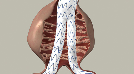
Radiology Waukesha has collaborated for 20 years with Vascular and Cardiothoracic Surgeon colleagues in the endovascular repair of thoracic aortic aneurysms and abdominal aortic aneurysms. Historically, abdominal aortic aneurysm treatment required major open abdominal surgery. Due to advances in the field, abdominal aortic aneurysms can now be treated by placing covered stents inside of the aorta through two small incisions in the groin. Even challenging cases such as aneurysms with complex necks can be treated in a minimally-invasive manner with the use of state-of-the-art techniques. With the goal of prevention, Radiology Waukesha offers abdominal aorta screening ultrasound, which provides early identification and ongoing monitoring of abdominal aortic aneurysms. Abdominal aortic aneurysms larger than 5 cm are at increased risk for rupture and will be promptly referred for appropriate treatment.
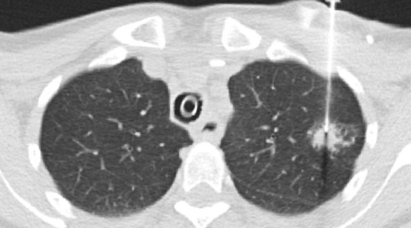
A biopsy removes a small sample of tissue from the body so the tissue can be examined by a pathologist. For most types of cancer, biopsy is considered the gold standard for obtaining a definitive cancer diagnosis. Radiology Waukesha radiologists are trained in minimally-invasive techniques which use image guidance to target even the smallest nodule or mass with exceptional accuracy and minimal risk. Imaging guidance varies by procedure and includes ultrasound, CT, MRI, and x-ray/fluoroscopy.
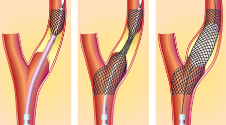
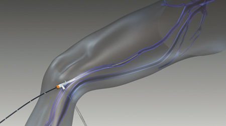
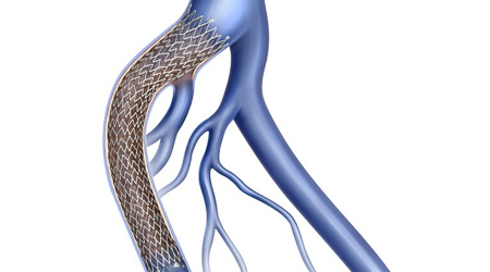
The carotid arteries are the main arteries that provide blood flow and oxygen to the brain. Plaque in the artery can build up (atherosclerosis), which causes severe narrowing of the carotid artery. To restore blood flow to the brain, the carotid artery needs to be opened up. This was traditionally achieved through an open surgical endarterectomy (artery clean out). However, certain patients are at high risk for stroke or adverse events from open surgery; therefore, open surgery is not always the ideal approach. An alternative to open surgery is endovascular carotid stent placement. In this minimally-invasive procedure, a small incision in the groin allows the Interventional Radiologist to pass a catheter up to the carotid artery in the neck. While using protection devices in the carotid artery, a stent is carefully deployed to restore blood flow to the brain. Radiology Waukesha has 20 years of experience in appropriate and safe endovascular carotid stent placement.
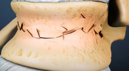
Osteopenia (reduced bone mass) and osteoporosis (bones are weak and brittle) are conditions that become more common with age. The loss of bone density puts patients at risk of breaking bones in their spine. These are called compression fractures, and they can be painful, can result in significant disability, and can be slow to heal. Radiology Waukesha Interventional Radiologists have extensive experience with minimally-invasive repair of broken vertebrae in the spine (vertebroplasty). Vertebroplasty is a minimally-invasive procedure which uses x-ray/fluoroscopic guidance to advance small needles into the broken bone. The fractures are “set” with various tools such as high-pressure balloons to restore bone height, and then the vertebral body is filled with "cement". Recovery is often immediate or within one week, accelerating the patient’s return to normal activity. In addition to compression fractures due to osteoporosis, broken bones related to spinal tumors can be treated using radiofrequency energy to destroy the tumor and then cement to heal the fracture.
Varicose veins can result in significant pain and morbidity. The veins in the legs have one-way valves which help keep the low pressure venous blood moving in one direction, up toward the heart. Some people (most commonly women, particularly women with prior pregnancy, and people with a family history of varicose veins) are at a greater risk for “leaky vein valves,” which results in blood pooling in the legs. This can result in leg swelling, varicose vein development, and in severe cases, non-healing ulcers around the ankle. Radiology Waukesha has a dedicated Vein Center with expertise in the diagnosis and treatment of varicose veins. The mainstay of treatment involves the minimally-invasive closure of these faulty veins using radiofrequency energy or “super glue” to close the vein shut. In addition, sclerotherapy may be offered, or microphlebectomy may be performed to remove visible large varicose veins. These procedures are all performed in the office and require little to no downtime for recovery.
Left leg swelling, pain, and large blood clots may form due to a common condition known as iliac vein compression syndrome (May-Thurner syndrome). This is a compression disorder of one of the large veins in the pelvis which results in internal scar tissue that slows blood flow from the left leg to the heart. Radiology Waukesha Interventional Radiologists have extensive experience in using x-ray/fluoroscopic guidance (venography) and intravascular ultrasound to assess the severity of the disease inside the vein. The narrowed vein is treated with a stent specifically designed to treat this medical problem through a tiny incision in the groin. This minimally-invasive procedure restores deep pelvic vein blood flow without a large incision or extended recovery time.